
Suji-gu, Yongin, Gyeonggi-do · SUJI VETERINARY CARDIOLOGY CENTER
Echocardiography for dogs and cats
If a murmur has been detected, or you want to assess your pet's condition while on heart medication, bring previous results and prescription details. We interpret ultrasound alongside history, auscultation, symptoms and other findings.
Why we review imaging and treatment records together
From examination findings
to treatment and follow-up decisions
Dr. Jinho Lee, DVM, MS, who completed a veterinary internal medicine residency focused on cardiology, assesses heart structure and blood flow. His experience includes medical care for refractory feline HCM heart failure, arrhythmias, cardiac tumors, severe heartworm infection and congenital heart disease complicated by endocrine or other conditions. Previous tests, medication and treatment responses inform the plan.
We explain current status and what to compare at the next visit. Before surgery or dental scaling at another clinic, you can discuss our cardiac assessment and report.
Clinical experience and academic activities · Managing concurrent cardiac and renal conditions
If a heart murmur has been detected
Tell us where the murmur was detected and whether previous tests or prescriptions are available. Auscultation alone does not determine the condition or treatment stage; examination and appropriate tests assess structure and blood flow.
Canine Myxomatous mitral valve disease (MMVD)and feline Hypertrophic cardiomyopathy (HCM), Congenital heart diseaseare assessed using individual findings and clinical status. Intermittent fainting or rhythm abnormalities may also warrant ECG or Holter monitoring.
For difficulty breathing, blue gums, fainting or sudden paralysis, go to the nearest 24-hour emergency veterinary hospital without waiting for an appointment or reply.
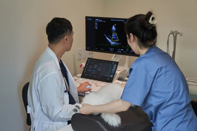
From examination to understanding results
- We review symptoms, previous images, reports and prescriptions, then explain the tests needed and estimated costs.
- We perform the agreed tests. Echocardiography assesses valves, chambers and blood flow, with interpretation alongside other findings as needed.
- Using images and measurements, we explain current status, treatment direction and changes to monitor.
For image copies and measurement reports included in detailed screening, see the health screening page. Before booking an individual test, ask about the records you need and how they can be provided.
For examination images and equipment capabilities, see our Samsung R20 equipment guide.
From echocardiography results to a treatment plan
We identify changes in the report and consider how they relate to current symptoms. The next decision varies with disease stage and previous treatment, even for the same diagnosis.
Canine valve disease: heart size and history matter
For MMVD, we assess valve leakage, left atrial and ventricular size, symptoms and any history of heart failure. Even without symptoms, criteria including heart size distinguish stages B1 and B2, affecting medication and follow-up decisions. Other indicated findings, including chest radiographs, are reviewed too.
Feline cardiomyopathy: wall thickness and the left atrium
When heart muscle appears thickened, we assess wall thickness, left atrial size and function, and blood flow. High blood pressure or hyperthyroidism can also change the heart, so further tests may help distinguish the cause. Left atrial changes inform assessment of heart failure and clotting risks and the follow-up plan.
Compare previous findings to decide what to check next
We compare test dates, images and prescriptions side by side, including the timing of medication changes and breathing or activity changes at home. If kidney values change during heart treatment, kidney function, electrolytes and blood pressure help determine the next checks.
When kidney values change during heart treatment · Care that reviews previous tests and prescriptions
Sources for condition-specific assessment: ACVIM canine valve disease consensus (2019) · ACVIM feline cardiomyopathy consensus (2020). Medication and recheck timing are determined individually during consultation.
Preparing your dog or cat for a visit
Bring previous reports, image files and medication packets or prescription photos. Names, doses and timing help us review treatment. Record videos of coughing or unsteadiness at home and note the circumstances.
Visiting with a dog
Tell us about pauses or coughing on walks and any changes after medication adjustments. Also tell us whether the visit is for routine assessment or new symptoms.
Visiting with a cat
Before booking, tell us about travel or clinic-related stress and current medications. We have a cat waiting area separate from dogs.
Anesthesia, sedation and fasting for echocardiography
Fasting, medication adjustments and sedation are advised individually according to health and the examination plan. Do not stop regular medication yourself; confirm the instructions you received for meals and medication.
View the full first visit checklist · Planning travel from southern Gyeonggi
Echocardiography fees and conditions
| Examination | Fee and conditions |
|---|---|
| Echocardiography | First visit KRW 200,000 / Follow-up KRW 100,000 After at least 1 health screening at our clinic: lifelong KRW 100,000 fee, regardless of weight or first/follow-up visit Interpretation included |
For consultation and other test fees, see our full fee guide. We explain recommended tests and estimated costs and perform agreed items. Confirm screening-history benefit eligibility before booking.
Visiting from Yongin or southern Gyeonggi Travel and parking by departure areaand Prepare previous results and medication detailsare available to help you prepare.
Planning heart care from southern Gyeonggi
The clinic is on the fourth floor of Suji Medipark, 65 Supung-ro, Suji-gu, Yongin. Regional guides cover the return journey, parking, previous records and medication details to bring.
Visitor guides for 21 cities & counties in southern Gyeonggi · Fees & applicable conditions · Hours & location