Skip to content
Veterinary resources
KakaoTalk ↗031-8067-7527Hours & location

Heart health checkups

SUJI Veterinary Cardiology Center, Yongin · Heart care for dogs & cats
Meet the veterinarian · Visit preparation · Test fees

Even if your pet seems well, you can discuss a checkup if you are concerned about heart health. We review everyday behavior and previous records to decide whether an assessment is needed.

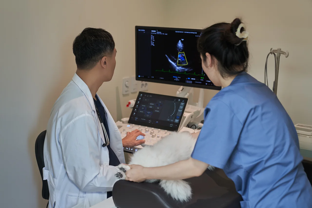
Dr. Lee Jinho performing heart ultrasound on a dog
Before deciding on a checkup

Start with the tests needed now

We consider a murmur, current heart medication and planned anesthesia alongside the history, auscultation and previous records to choose the timing and tests.

We do not perform every test at once. We explain why each is needed and its estimated cost, then proceed with your consent. Bring previous results and medication details.

What we consider together
  • Echocardiography is a key test of heart structure and blood flow. We consider the history, auscultation and necessary rhythm or imaging studies together to assess disease and its stage.
  • Changes in images and measurements help assess medication response and whether the disease stage has changed.
  • ECG, X-rays and echocardiography help review the evidence supporting the diagnosis.
  • We explain medical terms in Korean and English while reviewing the images with you.
  • We consider kidney, liver and electrolyte status alongside the heart. Medication is reduced when appropriate.
  • We offer pre-anesthetic cardiac assessment to help reduce anesthetic risk.
Who may benefit

Who may benefit

Checkup timing is based on the patient's condition and previous records. Cardiac assessment may be considered in the following situations.

  • Small breeds prone to MMVD — discuss timing based on breed, family history and routine auscultation, rather than age alone
  • Large breeds predisposed to DCM — discuss screening based on breed, family history, symptoms and auscultation
  • Cats with known Maine Coon or Ragdoll genetic variants, other breed predispositions, family history, breeding plans or abnormal auscultation
  • Any pet previously found to have a heart murmur
  • Older pets facing dental scaling or another procedure requiring anesthesia
  • Pets taking heart medication who need regular reassessment
  • Pets previously advised only to keep monitoring
  • Pets with another chronic condition, such as kidney or endocrine disease, needing coordinated care
What we check, and why

What each test shows

The heart and other organs affect each other, but not every patient needs the same tests. Below are ten options considered after reviewing the history, examination, auscultation and previous records. We explain the reasons and estimated costs before testing.

1. Blood tests

Assessing overall health and suitability for heart medication

Complete blood countCBC · Complete Blood Count

Why it may be needed

With anemia, the heart must beat faster and harder to carry oxygen around the body, adding strain to an already affected heart. Infection or inflammation can also cause signs resembling heart disease. This test helps assess the conditions in which the heart is working.

What it measures

Red blood cells (RBC), hemoglobin (Hb), hematocrit (HCT), white blood cells (WBC) and their types, and platelet count.

Interpreting the results
  • Low RBC / HCT — Indicates anemia. The additional workload on the heart must be considered when interpreting cardiac findings.
  • High RBC / HCT — May reflect dehydration, stress or excitement. Diuretic doses are reviewed if the patient takes them. Rarely, low oxygen from congenital heart disease increases red cell production; this is also considered in young patients.
  • High white cell count — Suggests infection or inflammation. We also consider whether coughing could be due to pneumonia rather than the heart.
  • Low platelet count — Prompts assessment of bleeding risk. However, Cavalier King Charles Spaniels naturally have larger, fewer platelets, and feline platelets may clump during sampling. A blood smear helps check whether the apparent low count is misleading.

Blood chemistrySerum Chemistry

Why it may be needed

Heart medications, especially diuretics, act through the kidneys. A drug that relieves the heart can therefore also affect the kidneys. Checking kidney and liver function before treatment helps assess tolerance. These parameters are monitored regularly after medication starts.

What it measures

We assess around 20 parameters, including liver enzymes (ALT, AST, ALP, GGT), total bilirubin, kidney markers (BUN, creatinine, SDMA), glucose, total protein, albumin, cholesterol and triglycerides.

Interpreting the results
  • High BUN / creatinine — Suggests kidney stress. Dehydration or reduced cardiac output can also raise these values, so they help guide diuretic adjustments. Urine concentration, measured as specific gravity, helps distinguish dehydration from intrinsic kidney disease.
  • Even with low creatinine — Kidney disease may still be present. Pets that lose muscle because of heart disease can have low creatinine despite impaired kidney function. SDMA, which is not affected by muscle mass, is also considered.
  • High liver enzymes — Can occur when right-sided heart failure causes blood to back up in the liver. Ultrasound helps distinguish a liver problem from a cardiac cause.
  • Low albumin — Makes fluid accumulation in the chest or abdomen more likely. It helps assess whether accumulated fluid is related to the heart.
  • High blood glucose — Prompts assessment for diabetes mellitus. Metabolic disease can change the overall cardiac treatment plan.

Example canine reference intervals: BUN 6–31 mg/dL · creatinine 0.5–1.6 mg/dL · ALT 12–121 U/L. Intervals vary by laboratory and equipment; use the reference intervals on the actual report.

ElectrolytesElectrolytes — Na · K · Cl

Why it may be needed

Electrical signals make the heart beat, and electrolytes help generate those signals. Potassium disturbances can cause dangerous arrhythmias. Diuretics can increase urinary potassium loss, so these values are checked particularly closely in pets taking heart medication.

What it measures

Blood sodium (Na), potassium (K), chloride (Cl), their balance and the Na/K ratio.

Interpreting the results
  • High potassium — May be associated with ACE inhibitors, spironolactone or impaired kidney function. It can slow the heart or cause dangerous arrhythmias, prompting immediate investigation and medication review.
  • Low potassium — Can be a diuretic adverse effect, causing weakness, reduced muscle strength and arrhythmias. We assess whether supplementation is needed.
  • Low sodium — May be seen with progressing heart failure and is considered when assessing prognosis.

Thyroid hormone testingThyroid Hormone — T4

Why it may be needed

The thyroid regulates metabolic pace. Excess hormone keeps the heart working faster and can thicken the heart muscle over time. Hyperthyroidism is a common cause of cardiac muscle thickening in older cats. Identifying it matters because thyroid treatment, rather than heart medication alone, addresses the cause.

What it measures

Measures total thyroxine (T4) in the blood. Free T4 is added when needed.

Interpreting the results
  • High T4 (mainly cats) — Raises suspicion of hyperthyroidism, which may cause a fast heart rate, weight loss and thickened heart muscle. Cardiac findings often improve when the thyroid condition is treated first.
  • Normal T4 (cats) — Does not completely exclude hyperthyroidism. Early disease or concurrent illness such as kidney disease can mask the elevation. An older cat with cardiac muscle thickening may need retesting in 2–4 weeks or additional free T4 testing.
  • Low T4 (mainly dogs) — May suggest hypothyroidism, associated with a slow heart rate, reduced energy and weight gain. Other chronic illnesses can lower T4 even with normal thyroid function, so T4 alone is not diagnostic. Compatible signs may prompt cTSH and free T4 testing.

2. Cardiac biomarkers

Looking for signals of cardiac stress before outward changes appear

NT-proBNPN-terminal pro B-type Natriuretic Peptide

Why it may be needed

NT-proBNP may rise when heart muscle is under pressure or stretched. It supports screening for possible structural heart disease and cardiac strain; a normal result cannot exclude mild or moderate disease. It is interpreted alongside symptoms, auscultation and kidney function, with echocardiography when abnormalities are suspected.

What it measures

Measures a circulating peptide reflecting the load or stress on heart muscle.

Interpreting the results
  • If elevated — Suggests greater cardiac strain. With symptoms, progression of heart disease is more likely, and echocardiography helps assess the cause and stage.
  • If within the reference interval — Suggests that cardiac strain may not be substantial at the time of testing, but does not exclude mild or moderate structural disease. The need for ultrasound depends on symptoms, auscultation and risk factors.
  • Keep in mind — Impaired kidney function can also raise the value, so kidney markers must be considered alongside it.

More about NT-proBNP results and further testing

Example canine interval: 0–900 pmol/L. The 900–1800 range is a gray zone; symptoms and ultrasound findings or repeat testing over time help interpretation. Cats have different thresholds, and units may be pmol/L or pg/mL depending on the assay.

Cardiac troponincTnI · cardiac Troponin I

Why it may be needed

Cardiac troponin I (cTnI) can rise with myocardial cell injury. It is selected and interpreted with symptoms, ECG and imaging when sudden fainting, unexplained arrhythmia or myocarditis is suspected. It can also support follow-up assessment in feline HCM.

What it measures

Measures the amount of a cardiac muscle contractile protein circulating in the blood.

Interpreting the results
  • If elevated — Suggests myocardial cell injury. We consider myocarditis, severe cardiomyopathy and injury associated with systemic illness. Short-interval repeat measurements may assess the trend. Myocardial infarction, common in people, is rare in dogs and cats.
  • If markedly elevated — Can indicate more extensive injury and a more guarded prognosis, helping guide treatment intensity.
  • If within the reference interval — Active myocardial injury may be less likely at present. Structural abnormalities are assessed separately by echocardiography.
  • Keep in mind — Troponin can also rise with non-cardiac conditions, including kidney dysfunction, severe systemic inflammation or sepsis, tumors, trauma and feline hyperthyroidism. It is interpreted with ultrasound and ECG findings, not by the number alone.

3. Imaging

Seeing structure and movement that numbers alone cannot show

Chest radiographyThoracic Radiograph — X-ray

Why it may be needed

Pulmonary edema, or fluid in the lungs, is a serious complication of heart disease. X-rays help identify it. Ultrasound shows the heart's structure; radiographs show the current lung changes associated with it. The tests complement rather than replace each other.

What it measures

Two or three views assess cardiac silhouette size (vertebral heart score, VHS), left atrial enlargement, pulmonary vessel size, lung patterns, tracheal elevation, left main bronchial compression and pleural fluid.

Interpreting the images
  • An enlarged cardiac silhouette — Indicates cardiac enlargement. The chambers involved help assess the type and progression of disease.
  • Increased whiteness in the lungs — May reflect pulmonary edema, pneumonia, hemorrhage or another cause. With left atrial enlargement, we assess the possibility of cardiogenic edema and congestive heart failure (CHF). Acute breathing difficulty, blue gums or fainting require the nearest 24-hour emergency veterinary hospital without waiting for a test appointment.
  • An enlarged left atrium displacing the bronchus — Prompts consideration of cardiac and airway causes of coughing. Left atrial enlargement alone often does not explain the cough, so lung patterns and airway findings are assessed together.
  • Clear lungs — Indicates no visible lung fluid at that time. The images provide a useful baseline for future comparison.

EchocardiographyEchocardiography

Why it may be needed

Direct assessment of cardiac structure, contraction, relaxation, valves and blood flow provides essential information for diagnosing and staging many structural diseases. History, examination, auscultation and relevant ECG, chest X-ray, blood pressure and blood test findings are also needed to assess rhythm and concurrent conditions.

General anesthesia is not used; most examinations are performed awake. If marked excitement prevents stable imaging, brief sedation is considered with the owner's agreement.

What it measures

Real-time images assess atrial and ventricular size, wall thickness, contraction, valve closure, and the direction and speed of blood flow using Doppler.

Interpreting the images
  • Thickened valves with backward blood flow — Consistent with MMVD. Left atrial and ventricular size (LA/Ao, LVIDDN), together with radiographic heart size (VHS), distinguish B1 from B2. Current or previous heart failure separates stages C and D. Medication starts at stage B2.
  • Thickened heart walls — Raises suspicion of HCM, the most common feline heart disease. Thrombus risk is assessed as well.
  • A dilated heart with reduced contraction — Consistent with DCM, seen mainly in large breeds. Early detection is particularly important.
  • Fluid in the sac around the heart — Indicates pericardial effusion. With low blood pressure, collapse or breathing difficulty, it may be compressing the heart, requiring immediate stabilization and treatment at the nearest 24-hour emergency veterinary hospital.
  • No structural abnormality — Some murmurs do not require treatment. We agree on when to reassess, rather than simply advising observation.

abdominal ultrasoundAbdominal Ultrasound

Why it may be needed

Owners often ask why the abdomen is scanned during a heart visit. When the heart cannot move blood forward effectively, it can back up in the liver and abdomen. A swollen belly cannot be identified as weight gain or fluid (ascites) by appearance alone. Kidney assessment also helps guide cardiac medication doses.

What it measures

We assess the liver, hepatic veins and caudal vena cava, abdominal fluid, kidney size and structure, and other abdominal organs including the spleen, adrenal glands and bladder.

Interpreting the images
  • Ascites with enlarged hepatic veins — Is common with right-sided heart failure. Pericardial effusion and liver or abdominal disease can also cause fluid, so echocardiography helps determine the cause before choosing a diuretic strategy.
  • Changes in kidney structure — Suggest concurrent chronic kidney disease (CKD). Heart and kidney treatment can place competing demands on each organ, requiring a balanced medication plan.
  • Enlarged adrenal glands — Prompts assessment for endocrine disease such as Cushing's syndrome, which can affect the heart through hypertension.
  • No other findings — Provides further support that the symptoms may be cardiac in origin.

4. Cardiac electrical activity

Assessing the rhythm, rather than simply the strength of the heartbeat

ECGECG · Electrocardiogram

Why it may be needed

Ultrasound assesses shape; ECG assesses rhythm. It is selected with the history and examination when arrhythmia may explain fainting or sudden weakness. Holter monitoring may be considered if a brief clinic recording is normal but suspicion remains.

What it measures

Records the rhythm and conduction of cardiac electrical signals as waveforms. We analyze P waves, QRS complexes, T waves, intervals, heart rate and regularity.

Interpreting the tracing
  • A very fast heartbeat — Is tachycardia. Waveforms help distinguish temporary clinic-related stress from an arrhythmia requiring treatment, such as atrial fibrillation.
  • A very slow heartbeat — Is bradycardia. If caused by conduction block, it can directly explain fainting.
  • Extra beats interrupting the rhythm — Are premature contractions. Their number and pattern help determine whether treatment is needed.
  • Symptoms despite a normal clinic ECG — May occur because the arrhythmia is intermittent. A Holter recording for 24 hours or longer can help identify the cause.
How we interpret the results. No single value establishes a diagnosis. Elevated NT-proBNP is interpreted differently when kidney markers also rise, and an enlarged cardiac silhouette can lead to different conclusions depending on ultrasound findings. History and examination define the clinical question; selected results are interpreted alongside age, symptoms and everyday behavior.
Step by step

Checkup steps & costs

The visit takes about 60–90 minutes. We review the images and explain the results with you the same day.

1 At check-in

History / respiratory-rate records

We review your sleeping respiratory-rate (SRR) records and videos of symptoms.

2 5–10 minutes

Cost estimate / consent

We explain today's proposed assessment and estimated cost first, then proceed with your consent.

3 10 minutes

Examination / auscultation / blood pressure

We assess murmur grade, breathing pattern and non-invasive blood pressure.

4 20–40 minutes

Selected tests — blood, imaging and ECG

History, examination and auscultation guide the selection and order of blood tests, chest X-rays, echocardiography, abdominal ultrasound and ECG. Every patient does not receive the same fixed set. We explain the reasons and estimated costs and perform the tests agreed to. Each test's role is described in the test guide below.

5 15–20 minutes

Results / home monitoring

We review images and measurements to explain stage and changes, and provide an SRR recording sheet.

Past promotion · September 28–October 2, 2026

Checkup fees

The opening discount ended on October 2, 2026. Before booking, call or message on KakaoTalk to confirm current checkup package prices and included tests. Individual test fees are listed in the Fee schedule.

View past opening promotion prices and conditions (September 28–October 2, 2026)
Past promotion only · not current prices
Checkup packageRegular price at the timePromotional price at the time
Comprehensive internal medicine checkup · all body weights at the timeKRW 1,074,000KRW 537,000
Comprehensive cardiac checkup · all body weights at the timeKRW 864,000KRW 432,000

Both packages were offered at 50% off the regular price. The quoted final amounts included D-dimer, urine protein:creatinine ratio (UPC), consultation and interpretation. Cortisol and total thyroxine (tT4) were separate tests at KRW 60,000 each. This record does not indicate current package prices or an extension of the discount.

An individual checkup plan

A heart checkup tailored to the patient

Tests are chosen using age, breed, symptoms, auscultation, clinical history and previous results. We discuss any tests needed beyond the checkup after the consultation.

Equipment note. The list shows tests that can be selected individually outside a checkup package. We explain the necessary items and estimated cost based on the consultation and proceed only with your consent.
Our approach to checkups

How do we approach checkups at SUJI?

More tests are not always necessary. We explain why a test is relevant to your pet, then review the findings together.

The same veterinarian throughout

The veterinarian performing the checkup discusses the results and sees your pet at the next visit, maintaining continuity of care.

Detailed images + clinical interpretation

Dr. Lee Jinho, a former veterinary internal medicine resident focused on cardiology, interprets Samsung R20 images and measurements alongside the examination and other test findings.

Imaging copies for you

A detailed checkup includes copies of the images and a measurement report for use during consultations at other hospitals.

A normal checkup still provides a plan

If no abnormalities are found, we explain when reassessment may be appropriate and record the suggested timing in the report.

Support for home monitoring

We provide an SRR measurement guide and recording sheet. If you notice concerning changes, contact us with the records.

Concurrent disease / multiple medications in older pets

We review heart, kidney, liver and endocrine status alongside prescriptions from other hospitals. Test findings and medication response guide whether drugs can be reduced or adjusted.

FAQs

FAQs

Does my pet need a checkup without symptoms?
Heart disease can progress without symptoms, but the same starting age does not apply to every pet. Breed, family history, breeding plans, murmurs, gallop sounds, arrhythmias, symptoms and previous results guide individual screening and recheck intervals.
Why are there so many tests? Are all of them necessary?
Each test provides different information about structure, rhythm, lungs or medication safety, although some findings overlap. We start with the history, examination, auscultation and previous records, then choose the fewest tests needed to answer the current question. Their roles are explained in “What these tests tell us”. We explain why each test is proposed and its estimated cost, and proceed with your consent.
Is fasting required?
Fasting requirements depend on the tests, age, underlying conditions and medications. Do not fast your pet on your own; follow the individual instructions provided when booking.
Can we hear the results the same day?
We review imaging and examination findings with you the same day. NT-proBNP and cardiac troponin I can also be tested in-house with Vcheck. We explain when results will be available according to the tests and progress.
Is anesthesia needed?
Most checkup tests do not require anesthesia. Brief sedation may help obtain more accurate images in highly excited patients or when otherwise needed. Anesthesia or sedation is discussed with you beforehand.
Can I bring images from another hospital?
Yes. CDs and photos are welcome. Comparing previous images helps assess change and can reduce duplicate tests.
Can another hospital use the report?
Yes. A detailed checkup includes imaging copies and a measurement report, prepared for use at another hospital, including during emergency care.
What is the total cost?
The opening discount ended on October 2. Ask before booking about current checkup package prices and included tests. Individual fees are in the Fee schedule. The lifetime echocardiography benefit for pets with a clinic checkup history is explained separately above.

Discuss a heart checkup

Tell us about your pet's usual behavior and your concerns. Bring previous test results if available.

Checkup guide

Could my pet benefit from a heart checkup?

The number of checked items cannot diagnose disease or determine severity. If even one item concerns you, discuss it at an appointment. Breathing difficulty, blue gums, fainting, sudden paralysis or severe pain require immediate assessment at the nearest 24-hour emergency veterinary hospital, regardless of the checklist result.

Breed information

Breed alone does not determine testing time

We discuss the necessary assessment after considering age, symptoms, auscultation and previous records.

Planning heart care from southern Gyeonggi

The clinic is on the fourth floor of Suji Medipark, 65 Supung-ro, Suji-gu, Yongin. Regional guides cover the return journey, parking, previous records and medication details to bring.

Visitor guides for 21 cities & counties in southern Gyeonggi · Fees & applicable conditions · Hours & location